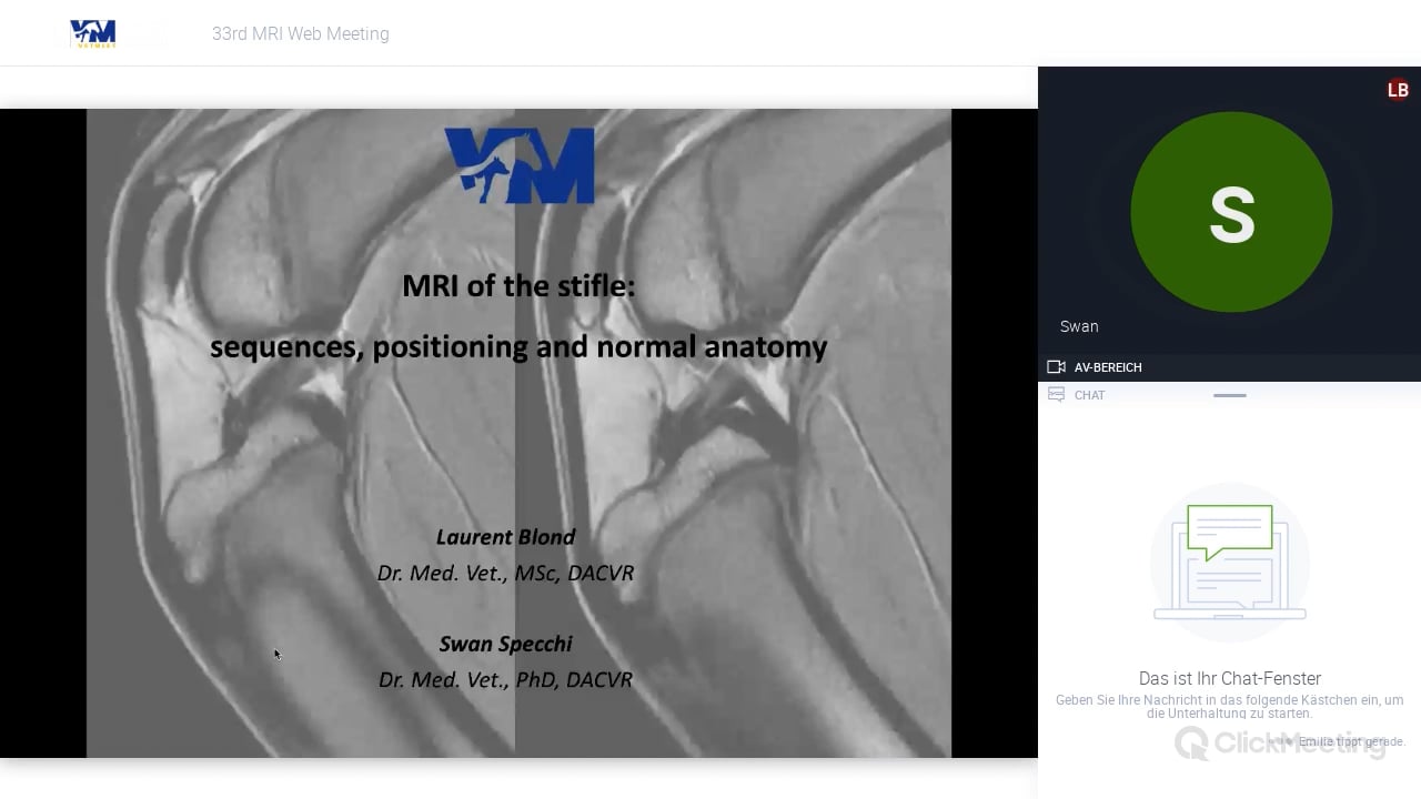
MRI of the stifle – sequences, positioning and normal anatomy
Description
Participants are expected to understand the common sequences used to image the stifle with MRI . Participants are expected to recognize the normal anatomy of the stifle imaged with MRI. This includes main bony structures, cartilage, synovial membrane, patellar and collateral ligaments and menisci.
Topics
Credits needed to watch:
1
Your current credits:
You need to log in to view your current credits. Login

 Hill's Vorbereitungskurs Ernährungsberatung
 Hill's Aufbaukurs - Ernährung des Hautpatienten


